


The Development History Of Veterinary X-ray Machines
Jan 27, 2026
Leave a message
In modern veterinary clinical practice, X-ray examination has become one of the basic diagnostic tools. Whether it is small animal diagnosis and treatment, health management of commercial animals, or sports medicine examination of large animals such as horses, veterinary X-ray machines play a key role in disease screening, skeletal assessment, and soft tissue evaluation. Looking back at its development history, veterinary X-ray machines have undergone continuous upgrades from "usable" to "easy to use," and then to "intelligent, efficient, and portable."

The beginnings of veterinary imaging (early 20th century)
After its discovery in 1895, X-ray technology was quickly applied to the medical field and gradually extended to the veterinary industry. Early veterinary X-ray machines were mostly modified from human equipment, making them bulky, complex to operate, and requiring extremely high environmental standards. Due to inaccurate exposure control and limited image clarity, X-ray examinations at this stage were mainly used for simple diagnoses such as animal fractures and foreign body ingestion, but they still opened the door to "visual diagnosis" for veterinary medicine.
Structural optimization and enhanced safety awareness (mid-to-late 20th century)
With the development of electronic technology, simulated X-ray machines specifically designed for veterinary clinical use gradually emerged. These machines began to have their power and exposure parameters adjusted for different animal sizes, significantly improving operational stability and imaging consistency. Simultaneously, the veterinary industry placed increasing emphasis on radiation protection, and protective clothing, collimators, and other supporting equipment became more widespread, making X-ray examinations safer and more standardized in clinical settings.
During this period, veterinary X-ray machines became essential fixed equipment in animal hospitals, but they were still primarily "wired powered and fixedly installed," limiting their flexibility.
DR systems drive a leap in efficiency (early 21st century)
In the 21st century, the introduction of digital imaging technology (CR, DR) has completely transformed the way veterinary X-ray machines are used. DR systems enable instant image display, significantly reducing examination time and improving diagnostic efficiency. Images can be magnified, measured, compared, and stored, facilitating long-term case management and remote consultations.
Simultaneously, digital technology has significantly improved image quality while reducing X-ray dose, making soft tissue and bone details clearer and providing a more reliable basis for the diagnosis of complex diseases.
Adapting to the needs of diagnosis and treatment in multiple scenarios (last decade)
In recent years, with the growing demand for mobile healthcare and large animal diagnosis, the development focus of veterinary X-ray machines has gradually shifted towards portability, wireless connectivity, and intelligence. The maturation of technologies such as rechargeable batteries, wireless transmission, and preset parameter programs has enabled X-ray examinations to move beyond fixed examination rooms and into ranches, horse farms, and field rescue sites.
Meanwhile, intelligent preset parameters tailored to different animal sizes and body parts significantly lower the operational threshold and improve the success rate of imaging, making it particularly suitable for veterinary applications with fast-paced work and diverse scenarios.
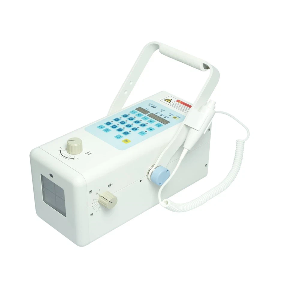
Against the backdrop of veterinary X-ray machines continuously evolving towards higher performance and greater flexibility, the x-ray machine for veterinary is a representative product of this development trend.
This device features a compact and lightweight design, making it easy to carry and deploy quickly. It can output excellent high-quality images in a variety of environments, making it an ideal choice for veterinary clinical practice and large animal examination.
Key advantages include
- High power adaptability: Supports 100~240V, suitable for different countries and regions.
- Powerful 90kV, 40mA output: Meets various animal examination needs.
- Rechargeable battery powered:Allows for 300 exposures on a full charge.
- Battery powered, no cables required, enabling truly wireless operation.
- DR system + one-stop image acquisition station:Capture, display, and management all in one step.
- Higher image quality, lower X-ray dose, balancing diagnostic effectiveness and safety.
- 32 preset parameters for 4 body types and 8 body parts:Enhanced operational efficiency.
- Two transportation options:Flight case and carry-on case, adaptable to different outpatient scenarios.
- Customized services supported:Meets individual clinical needs.
Of particular note is the exceptional suitability of this veterinary X-ray machine for equine examination and diagnosis. Whether assessing limb skeletal structure or screening for sports injuries, it enables rapid, high-quality imaging on-site.
From its initial analog imaging capabilities to today's digital, portable, and intelligent versions, the history of veterinary X-ray machines reflects the ever-increasing precision and efficiency in veterinary medicine. In the future, with the further integration of artificial intelligence and telemedicine technologies, veterinary imaging equipment will continue to evolve, providing even stronger technical support for animal health management. And next-generation products like the x-ray machine for veterinary are becoming indispensable and reliable partners for modern veterinarians.
Send Inquiry